What is it?
Spectroscopy and spectrometry let chemists identify a compound without a chemical test, using only a few milligrams.
- Infrared (IR) spectroscopy shines infrared light through a sample. Covalent bonds vibrate (stretch and bend), and each type of bond absorbs IR at a characteristic wavenumber, measured in cm⁻¹. The absorptions show which functional groups are present.
- Mass spectrometry (MS) turns molecules into positive ions, breaks some of them into fragments, and sorts the ions by their mass-to-charge ratio, . The heaviest ion usually gives the molar mass.
The wavenumber is the number of waves per centimetre, the reciprocal of the wavelength: . A higher wavenumber means a higher frequency and more energy.
Key idea
IR answers “which functional groups?”; mass spectrometry answers “how heavy, and in what pieces?” Together with NMR, they let chemists work out the structure of almost any organic compound.
Why does it matter?
- Identifying unknowns. Forensic laboratories, drug testing and quality control identify substances from their spectra.
- Breathalysers measure ethanol in breath using its IR absorption.
- Climate. Carbon dioxide, methane and water vapour absorb infrared radiation from the Earth, which is the greenhouse effect.
- Space and medicine. Mass spectrometers analyse the atmospheres of planets, detect drugs in sport and identify proteins in disease.
How does it work?
1. Reading an IR spectrum
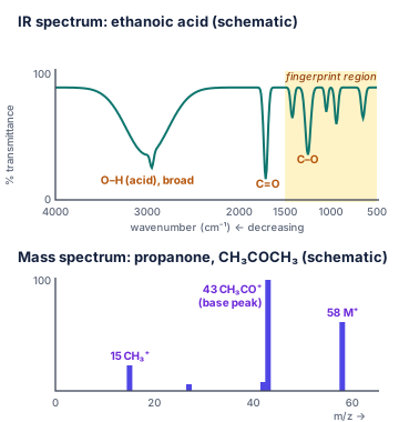
An IR spectrum plots transmittance (%) against wavenumber, with wavenumber decreasing from left (about 4000 cm⁻¹) to right (about 500 cm⁻¹). Absorptions appear as dips (“peaks” pointing down).
| Bond | Where found | Wavenumber (cm⁻¹) | Appearance |
|---|---|---|---|
| O–H | alcohols | 3200–3550 | strong, broad |
| O–H | carboxylic acids | 2500–3300 | very broad |
| N–H | amines, amides | 3300–3500 | medium |
| C–H | almost all organic compounds | 2850–3100 | medium–strong |
| C≡N | nitriles | about 2250 | medium, sharp |
| C=O | aldehydes, ketones, acids, esters | 1680–1750 | strong, sharp |
| C=C | alkenes | 1620–1680 | weak–medium |
| C–O | alcohols, esters, ethers | 1000–1300 | strong |
The region below about 1500 cm⁻¹ contains many overlapping absorptions. It is called the fingerprint region: it is unique to each compound, so it can be matched against a database to confirm an identity.
2. Reading a mass spectrum
A mass spectrum is a bar chart of relative abundance against (for ions with a 1+ charge, is simply the mass).
- The molecular ion, , is the peak at the highest (ignoring small isotope peaks). Its equals the molar mass.
- The tallest peak is the base peak (set to 100 %). It comes from the most stable fragment.
- Fragments reveal pieces of the molecule: 15 (), 29 ( or ), 31 (), 43 ( or ), 45 ().
- Isotope patterns: a compound with one Cl atom shows peaks at M and M+2 in a 3 : 1 ratio ( and ); one Br atom gives M and M+2 in a 1 : 1 ratio.
Think of it like this
IR spectroscopy is like listening to an orchestra: each type of bond “plays” at its own pitch, so you can hear which instruments (functional groups) are present. Mass spectrometry is like weighing a vase and then the pieces it breaks into: the whole vase gives the total mass, and the pieces hint at how it was put together.
More precisely
A bond absorbs IR only if its vibration changes the dipole moment of the molecule, which is why and do not absorb but and do. Stronger bonds and lighter atoms vibrate faster, so they absorb at higher wavenumbers: C≡C absorbs higher than C=C, which absorbs higher than C–C. In MS, the most common method (electron impact) knocks an electron out of the molecule; the small M+1 peak comes mainly from carbon-13 (1.1 % of carbon atoms), and high-resolution MS measures masses to four decimal places, enough to fix the molecular formula.
Visualise it
Worked example
Worked example: Wavenumber and wavelength
Question: A C=O absorption appears at 1715 cm⁻¹. What is its wavelength in micrometres?
Worked example: Alcohol, acid or ketone?
Question: Three compounds give these IR absorptions. Identify the functional group in each. (a) broad 3350 cm⁻¹, 1050 cm⁻¹, nothing near 1700 cm⁻¹ (b) very broad 2500–3300 cm⁻¹ and strong 1710 cm⁻¹ (c) strong 1715 cm⁻¹, no absorption above 3100 cm⁻¹
- (a) O–H (alcohol) and C–O, no C=O: an alcohol.
- (b) Very broad acid O–H plus C=O: a carboxylic acid.
- (c) C=O but no O–H: a ketone or aldehyde (or ester).
Worked example: Average atomic mass from a mass spectrum
Question: The mass spectrum of chlorine atoms shows (34.969, 75.76 %) and (36.966, 24.24 %). Calculate the relative atomic mass of chlorine.
The 3 : 1 ratio of the isotopes is why chlorine compounds show M and M+2 peaks in a 3 : 1 ratio.
Worked example: Putting IR and MS together
Question: An unknown has the formula . Its IR spectrum shows a strong absorption at 1715 cm⁻¹ and none at 3200–3550 cm⁻¹. Its mass spectrum shows at 58 and the base peak at 43. Identify it.
- = 58 matches (58.08 g/mol).
- IR: C=O present, no O–H: an aldehyde or a ketone.
- MS: a base peak at 43 is , formed when the molecule loses (58 − 15 = 43).
- The compound is propanone, . (Its isomer propanal would give a strong peak at 29, or .)
Common mistake
Common mistake: Reading the wavenumber axis the wrong way
IR spectra are drawn with wavenumber decreasing from left to right. An O–H band near 3300 cm⁻¹ is on the left; C=O near 1700 cm⁻¹ is in the middle.
Common mistake: Taking the tallest peak as the molar mass
The tallest peak is the base peak, usually a fragment. The molar mass is given by the molecular ion, the peak at the highest (apart from small isotope peaks).
Common mistake: Trying to identify everything in the fingerprint region
Below 1500 cm⁻¹ the absorptions overlap. Use the fingerprint region to match a known spectrum, and the region above 1500 cm⁻¹ to identify functional groups.
Notation note
- Wavenumber: , in cm⁻¹ (read “per centimetre” or “reciprocal centimetres”).
- : molecular ion; M+2: an ion 2 mass units heavier, usually from an isotope.
Remember this
Remember this
- IR: bonds absorb at characteristic wavenumbers. Broad 3200–3550: alcohol O–H. Very broad 2500–3300: acid O–H. Strong 1680–1750: C=O.
- Below 1500 cm⁻¹: fingerprint region, unique to each compound.
- MS: molecular ion (highest m/z) = molar mass; base peak = tallest; fragments show pieces.
- Cl: M and M+2 in 3 : 1; Br: 1 : 1.
- Combine: MS gives the mass, IR the functional groups, NMR the carbon–hydrogen framework.
Test yourself
Check your understanding before moving on.
Flashcards
IR Spectroscopy and Mass Spectrometry: Flashcards
- QuestionWhat does an IR spectrum show?Answer
Which bonds (functional groups) are present: each bond absorbs IR at a characteristic wavenumber.
- QuestionWhat is a wavenumber?Answer
The number of waves per centimetre, 1/λ, in cm⁻¹. Higher wavenumber = higher energy.
- QuestionWhere does a C=O bond absorb in IR, and what does it look like?Answer
1680–1750 cm⁻¹, strong and sharp.
- QuestionHow do the O–H absorptions of alcohols and carboxylic acids differ?Answer
Alcohol: broad, 3200–3550 cm⁻¹. Acid: very broad, 2500–3300 cm⁻¹.
- QuestionWhat is the fingerprint region?Answer
Below about 1500 cm⁻¹: complex, unique to each compound, used to match a known spectrum.
- QuestionIn a mass spectrum, what is the molecular ion?Answer
M⁺, the peak at the highest m/z (ignoring small isotope peaks); its m/z equals the molar mass.
- QuestionWhat is the base peak?Answer
The tallest peak (100 %), from the most stable fragment.
- QuestionGive the ions for fragments at m/z 15, 29 and 43.Answer
15: CH₃⁺. 29: C₂H₅⁺ or CHO⁺. 43: C₃H₇⁺ or CH₃CO⁺.
- QuestionWhat M : M+2 ratio shows one Cl atom? One Br atom?Answer
Cl: 3 : 1. Br: 1 : 1.
- QuestionWhy do N₂ and O₂ not absorb IR?Answer
Their vibration does not change the dipole moment of the molecule.
Tip: press Space to flip and ← → to move between cards.
Quiz
IR Spectroscopy and Mass Spectrometry: Quiz
7 questions
C=O bonds absorb strongly at 1680–1750 cm⁻¹ (aldehydes, ketones, acids, esters).
Show answer
Answer: C=O
C=O bonds absorb strongly at 1680–1750 cm⁻¹ (aldehydes, ketones, acids, esters).
A broad band at 3200–3550 cm⁻¹ is an alcohol O–H. No C=O rules out ketones and acids.
Show answer
Answer: an alcohol
A broad band at 3200–3550 cm⁻¹ is an alcohol O–H. No C=O rules out ketones and acids.
The molecular ion M⁺ is the whole molecule minus one electron; its m/z gives the molar mass. The base peak is the tallest peak.
Show answer
Answer: molecular ion
The molecular ion M⁺ is the whole molecule minus one electron; its m/z gives the molar mass. The base peak is the tallest peak.
³⁵Cl and ³⁷Cl occur in about a 3 : 1 ratio, giving M and M+2 peaks 2 units apart in that ratio. One Br would give 1 : 1.
Show answer
Answer: One chlorine atom
³⁵Cl and ³⁷Cl occur in about a 3 : 1 ratio, giving M and M+2 peaks 2 units apart in that ratio. One Br would give 1 : 1.
58 − 43 = 15: loss of a methyl group leaves CH₃CO⁺ at m/z 43.
Show answer
Answer: CH₃ (15)
58 − 43 = 15: loss of a methyl group leaves CH₃CO⁺ at m/z 43.
λ = 1/3300 cm⁻¹ = 3.03 × 10⁻⁴ cm = 3.03 μm (1 cm = 10⁴ μm).
Show answer
Answer: 3.03 μm
λ = 1/3300 cm⁻¹ = 3.03 × 10⁻⁴ cm = 3.03 μm (1 cm = 10⁴ μm).
A carboxylic acid has both an O–H (very broad, overlapping the C–H region) and a C=O. The first pair is an alcohol; 2250 is C≡N; 1650 + 3050 suggests an alkene.
Show answer
Answer: Very broad 2500–3300 cm⁻¹ and strong 1700–1725 cm⁻¹
A carboxylic acid has both an O–H (very broad, overlapping the C–H region) and a C=O. The first pair is an alcohol; 2250 is C≡N; 1650 + 3050 suggests an alkene.
Notes and downloads
Worksheet
IR Spectroscopy and Mass Spectrometry Worksheet
8 questions on IR absorptions and functional groups, wavenumbers, molecular ions, fragments, isotope patterns and identifying an unknown. Answer key included.
References
- Brown, T. L.; LeMay, H. E., Jr.; Bursten, B. E.; Murphy, C. J.; Woodward, P. M.; Stoltzfus, M. W. Chemistry: The Central Science, 15th ed.; Pearson, 2022.
Practise this topic with flashcards and a quiz at chemistryclarity.com/chemistry/ir-and-mass-spectrometry/
Spotted a mistake? Let us know and we'll fix it.