What is it?
Radiation from radioactive nuclei is ionizing: it has enough energy to knock electrons out of atoms and molecules in the body. This can break bonds in DNA directly, or split water into reactive free radicals that damage cells. The same property makes radiation dangerous and useful: it can harm healthy tissue, but it can also be aimed at cancer cells, and the gamma rays that escape the body can be detected to make images.
Three quantities describe radiation and its effects:
| Quantity | Unit | Meaning |
|---|---|---|
| Activity | becquerel, Bq | 1 Bq = 1 decay per second |
| Absorbed dose | gray, Gy | 1 Gy = 1 J of energy absorbed per kg of tissue |
| Equivalent dose | sievert, Sv | absorbed dose × radiation weighting factor |
The radiation weighting factor is 1 for gamma rays, X-rays and beta particles, but 20 for alpha particles, which deposit their energy in a very short track and do much more damage per joule.
Key idea
Medicine uses the right radiation for the job. Imaging needs gamma rays that escape the body, from an isotope with a short half-life. Therapy needs radiation that deposits its energy locally (beta or alpha particles), or a carefully aimed external beam.
Why does it matter?
- Diagnosis. Millions of scans a year use technetium-99m to image bones, the heart and other organs, and fluorine-18 PET scans find cancers and study the brain.
- Treatment. Iodine-131 treats overactive and cancerous thyroid glands; external gamma and X-ray beams and implanted sources treat many cancers.
- Safety. Patients, staff and the public must be protected, so doses are measured, limited and kept as low as reasonably achievable (ALARA).
How does it work?
1. Choosing a tracer for imaging
A good imaging isotope:
- emits gamma rays (they pass out of the body to the camera; alpha and beta would be absorbed and only cause damage);
- has a short half-life (hours), long enough for the scan but short enough to limit the dose;
- can be attached to a molecule that collects in the organ of interest.
Technetium-99m ( = 6.01 h, 140 keV gamma rays) fits all three. Iodine-123 ( = 13.2 h) is taken up by the thyroid, which uses iodine.
2. PET scans
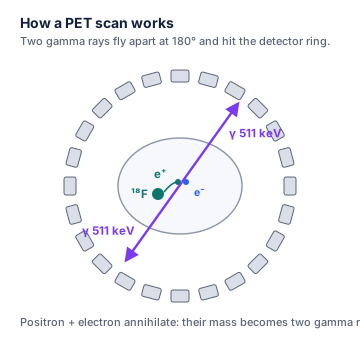
In positron emission tomography, a glucose-like molecule labelled with fluorine-18 ( = 110 min) collects in very active tissue such as tumours. Each fluorine-18 nucleus emits a positron, which meets an electron within a millimetre or so. The two annihilate, and their mass becomes two 511 keV gamma rays that fly apart in opposite directions. Detectors around the patient record both, and a computer traces back where they came from.
3. Radiotherapy
- Iodine-131 (beta and gamma emitter, = 8.02 d) concentrates in the thyroid, where its beta particles destroy the tissue.
- External beams of high-energy X-rays or gamma rays are aimed at a tumour from several directions, so the tumour gets a high dose while surrounding tissue gets less.
- Brachytherapy places small sealed sources inside or next to the tumour. Some newer treatments use alpha emitters, such as radium-223 ( = 11.4 d) for cancer that has spread to bone.
4. Protection: time, distance, shielding
- Time: dose = dose rate × time, so spend as little time near a source as possible.
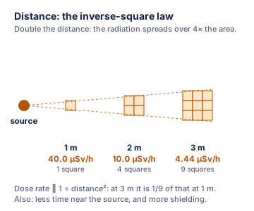
- Distance: the dose rate from a small source falls with the square of the distance (inverse-square law): .
- Shielding: lead, concrete or water absorbs radiation. Each half-value thickness of a shield halves the intensity.
Workers wear dosimeters that record their dose, and dose limits are set well below the levels known to cause harm.
| Dose (equivalent) | Example |
|---|---|
| about 2.4 mSv per year | worldwide average natural background (radon, rocks, cosmic rays, food) |
| 1 mSv per year | limit for the public from artificial sources (excluding medical) |
| 20 mSv per year | limit for radiation workers (averaged over 5 years) |
Think of it like this
Standing near a radioactive source is like standing near a campfire. You warm up less if you stay a shorter time (time), step back (distance: much cooler two steps away), or stand behind a wall (shielding). The fire itself (the activity) stays the same; what changes is how much reaches you.
More precisely
The effective dose (also in sieverts) goes one step further: it weights each organ by its sensitivity to radiation, so that doses to different parts of the body can be compared and added. In a patient, the activity of a tracer falls both by radioactive decay and by the body excreting it; the combined rate is described by an effective half-life, which is shorter than the physical half-life. Radiation effects are of two kinds: high doses cause predictable tissue damage (such as burns or radiation sickness), while low doses slightly raise the long-term probability of cancer, which is why every unnecessary dose is avoided.
Visualise it
Worked example
Worked example: Absorbed dose and equivalent dose
Question: A 70.0 kg person absorbs 0.0150 J from gamma rays. Find the absorbed dose and the equivalent dose. What would the equivalent dose be if the same energy came from alpha particles?
- Absorbed dose
- Gamma (weighting factor 1): 0.214 mSv
- Alpha (weighting factor 20): 4.29 mSv, twenty times more harmful for the same energy.
Worked example: Distance and the inverse-square law
Question: The dose rate 1.00 m from a source is 40.0 µSv/h. What is it at 2.00 m and at 3.00 m?
- At 2.00 m: 10.0 µSv/h
- At 3.00 m: 4.44 µSv/h
Worked example: How much tracer is left?
Question: A patient receives 740 MBq of technetium-99m ( = 6.01 h). What activity remains 18.0 h later (ignoring excretion)?
- half-lives
- 92.8 MBq, about one-eighth of the starting activity.
Common mistake
Common mistake: Confusing activity with dose
A source with a high activity (Bq) does not necessarily give a high dose: the dose depends on the type and energy of the radiation, how much is absorbed, the distance and the time. Activity describes the source; dose describes what the body receives.
Common mistake: Halving the dose rate when the distance doubles
The inverse-square law means doubling the distance divides the dose rate by 4, not 2; tripling it divides by 9.
Common mistake: Thinking an imaged patient becomes permanently radioactive
Tracers have short half-lives and are also excreted, so the activity falls quickly. After a technetium-99m scan, the activity is about one-sixteenth of the starting value within a day. External X-ray and gamma beams leave no radioactivity in the body at all.
Notation note
- Prefixes: 1 mSv = 10⁻³ Sv; 1 µSv = 10⁻⁶ Sv; 1 MBq = 10⁶ Bq.
- Older units: 1 curie (Ci) = 3.7 × 10¹⁰ Bq; 1 rad = 0.01 Gy; 1 rem = 0.01 Sv.
- keV and MeV are units of energy for single photons or particles (1 keV = 1.602 × 10⁻¹⁶ J).
Remember this
Remember this
- Activity in Bq (decays per second); absorbed dose in Gy (J/kg); equivalent dose in Sv = Gy × weighting factor (1 for γ, X, β; 20 for α).
- Imaging: gamma emitters with short half-lives (Tc-99m, 6.01 h). PET: F-18 positrons give two 511 keV gamma rays.
- Therapy: beta or alpha emitters placed in the tumour (I-131, Ra-223), or aimed external beams.
- Protection: less time, more distance (rate ∝ 1/d²), more shielding; keep doses as low as reasonably achievable.
Test yourself
Check your understanding before moving on.
Flashcards
Nuclear Medicine and Radiation Safety: Flashcards
- QuestionWhat is ionizing radiation, and how does it harm cells?Answer
Radiation that knocks electrons out of atoms. It breaks bonds in DNA directly or forms damaging free radicals from water.
- QuestionBecquerel, gray, sievert?Answer
Bq = decays per second (activity). Gy = J absorbed per kg (absorbed dose). Sv = Gy × weighting factor (equivalent dose).
- QuestionRadiation weighting factors?Answer
1 for gamma, X-rays and beta; 20 for alpha.
- QuestionWhat makes a good imaging isotope?Answer
Gamma emitter, short half-life (hours), can be targeted to an organ. Example: technetium-99m (6.01 h).
- QuestionWhy are alpha emitters not used for imaging?Answer
Alpha particles do not leave the body, so they cannot be detected outside, and they give a high local dose.
- QuestionHow does a PET scan detect fluorine-18?Answer
Its positron annihilates with an electron, giving two 511 keV gamma rays in opposite directions, recorded by a detector ring.
- QuestionName a radioisotope used to treat the thyroid.Answer
Iodine-131 (beta and gamma, 8.02 d): the thyroid takes up iodine, and the beta particles destroy the tissue.
- QuestionThree ways to reduce radiation dose?Answer
Less time, more distance, more shielding (and keep doses as low as reasonably achievable).
- QuestionInverse-square law?Answer
Dose rate ∝ 1 ÷ distance²: doubling the distance cuts the rate to one-quarter.
- QuestionAverage natural background dose?Answer
About 2.4 mSv per year worldwide, mostly from radon, rocks, cosmic rays and food.
Tip: press Space to flip and ← → to move between cards.
Quiz
Nuclear Medicine and Radiation Safety: Quiz
7 questions
1 Gy = 1 J/kg. The becquerel measures activity; the sievert adds the radiation weighting factor.
Show answer
Answer: gray
1 Gy = 1 J/kg. The becquerel measures activity; the sievert adds the radiation weighting factor.
Gamma rays leave the body to reach the camera, and a short half-life limits the dose. This describes technetium-99m.
Show answer
Answer: a gamma emitter with a half-life of 6 hours
Gamma rays leave the body to reach the camera, and a short half-life limits the dose. This describes technetium-99m.
Equivalent dose = 0.010 Gy × 20 = 0.20 Sv.
Show answer
Answer: 0.20 Sv
Equivalent dose = 0.010 Gy × 20 = 0.20 Sv.
36 µSv/h × (1.0 m ÷ 3.0 m)² = 36 µSv/h ÷ 9 = 4.0 µSv/h.
Show answer
Answer: 4.0 µSv/h
36 µSv/h × (1.0 m ÷ 3.0 m)² = 36 µSv/h ÷ 9 = 4.0 µSv/h.
Positrons annihilate with electrons inside the body, producing pairs of 511 keV gamma rays at 180°.
Show answer
Answer: two gamma rays travelling in opposite directions
Positrons annihilate with electrons inside the body, producing pairs of 511 keV gamma rays at 180°.
6.0 mm ÷ 2.0 mm = 3 half-value thicknesses, so (½)³ = 1/8 gets through.
Show answer
Answer: 1/8
6.0 mm ÷ 2.0 mm = 3 half-value thicknesses, so (½)³ = 1/8 gets through.
Chemistry delivers the isotope to the right organ; the short-range beta particles deposit their energy there.
Show answer
Answer: the thyroid takes up iodine, and its beta particles destroy nearby tissue
Chemistry delivers the isotope to the right organ; the short-range beta particles deposit their energy there.
Notes and downloads
Worksheet
Nuclear Medicine and Radiation Safety Worksheet
9 questions on radiation units, choosing isotopes, dose calculations, the inverse-square law, shielding and PET. Answer key included.
References
- Brown, T. L.; LeMay, H. E., Jr.; Bursten, B. E.; Murphy, C. J.; Woodward, P. M.; Stoltzfus, M. W. Chemistry: The Central Science, 15th ed.; Pearson, 2022.
Practise this topic with flashcards and a quiz at chemistryclarity.com/chemistry/nuclear-medicine/
Spotted a mistake? Let us know and we'll fix it.